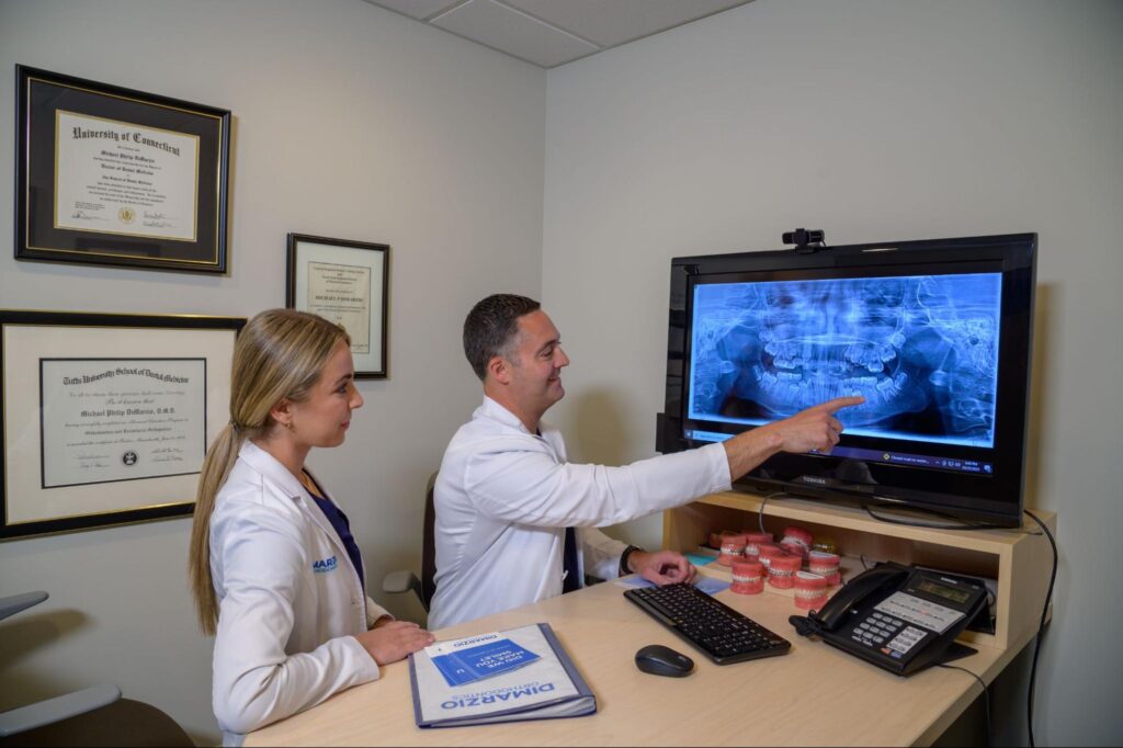
Every orthodontic journey at DiMarzio Orthodontics begins with an X-ray, the unseen guide mapping the path to a perfectly aligned smile. Under the expert eyes of Dr. DiMarzio and Dr. Marczak, these powerful diagnostic images reveal the hidden contours of your dental structure, setting the stage for precision in every adjustment. Join us as we explore how X-rays provide the foundation for personalized treatment plans that transform smiles with meticulous accuracy.
X-Rays in Orthodontic Diagnosis
At DiMarzio Orthodontics, X-rays are vital for diagnosing and crafting precise orthodontic treatments. These diagnostic images reveal much more than what meets the eye, providing a deep view into the hidden structures of the mouth. Here’s how they support our expert team:
- Uncovering Hidden Details: X-rays expose the roots, bones, and hidden aspects of dental anatomy, essential for a comprehensive health assessment.
- Identifying Orthodontic Issues: They pinpoint issues like misalignments and overcrowding, crucial for early and effective intervention.
- Customizing Treatment Plans: With detailed insights from X-rays, Dr. DiMarzio and Dr. Marczak tailor treatment plans to address each patient’s specific alignment needs.
X-rays are foundational at the outset and continue to guide adjustments throughout the treatment process. Next, we will explore how these images monitor progress and ensure treatments adapt to deliver optimal outcomes.

Tracking Your Smile’s Transformation with X-Rays
Continuous evaluation is crucial in orthodontics, and at DiMarzio Orthodontics, X-rays are integral to monitoring the progression of treatment. These periodic snapshots provide Dr. DiMarzio and Dr. Marczak with real-time updates on how well teeth are moving and adapting to braces or aligners. Here’s how X-rays contribute to ensuring successful outcomes:
Tracking Tooth Movement
X-rays show detailed views of tooth positions, allowing for precise adjustments. This ensures that the movement of each tooth is progressing according to the treatment plan.
Assessing Bone Response
Beyond just teeth, X-rays help evaluate how the jawbone is responding to the forces applied by orthodontic devices. This information is vital to prevent any long-term damage and to make necessary modifications to the treatment.
Guiding Adjustments
As the treatment progresses, X-rays provide the evidence needed to make informed decisions about whether to continue the current course or to make adjustments. This adaptability helps in achieving the desired end result more efficiently.
With the help of these detailed X-rays, every step in the orthodontic process is informed and exact. In the next section, we’ll discuss the technological advancements in X-ray imaging that enhance the treatment process at DiMarzio Orthodontics.
Technological Advancements in X-Ray Imaging
At DiMarzio Orthodontics, we leverage cutting-edge X-ray technology to enhance the precision and safety of our orthodontic treatments. These technological advancements have revolutionized how we view and utilize X-rays, providing clearer images with less exposure to radiation. Here’s how these innovations benefit our practice and patients:
Digital X-Ray Technology
This modern approach reduces radiation exposure significantly compared to traditional methods while delivering higher-quality images almost instantaneously. Digital X-rays allow us to assess fine details and make more accurate assessments.
3D Imaging
Utilizing three-dimensional X-ray views gives us a comprehensive understanding of the patient’s craniofacial structure. This depth of detail is crucial for complex cases involving jaw alignment and other structural considerations, ensuring that our treatments are both effective and tailored to individual needs.
Enhanced Patient Communication
These advanced imaging technologies not only aid in diagnosis and treatment but also enhance communication with our patients. We can show clear, understandable images, explaining the issues and outlining the treatment process in a way that is easy to grasp.
By integrating these advanced diagnostic tools, DiMarzio Orthodontics ensures that every treatment decision is based on detailed, accurate information. Next, we will discuss how we prepare patients for an X-ray procedure and what they can expect during their visit, making the experience as comfortable and informative as possible.
Preparing for an X-Ray at DiMarzio Orthodontics
At DiMarzio Orthodontics, we ensure our patients are well-prepared and comfortable before undergoing an X-ray. Here’s what to expect during your visit:
- Before the X-Ray: It’s important to remove any jewelry or metal accessories that might interfere with the image quality. Wearing comfortable clothing will also facilitate an easier process.
- During the X-Ray Procedure: Our technicians will guide you on how to position yourself, whether sitting or standing, to capture the most effective images. Safety is prioritized, so protective gear such as a lead apron will be provided to minimize radiation exposure.
- Reviewing Your X-Rays: Post-procedure, Dr. DiMarzio or Dr. Marczak will review the images with you, discussing any notable findings and how they impact your treatment plan.
This preparation and review process helps demystify the experience, making patients more comfortable and informed about their orthodontic care. Next, we’ll detail the safety measures implemented during X-ray procedures to ensure every patient’s well-being.
Ensuring Safety During X-Ray Procedures
At DiMarzio Orthodontics, patient safety during X-ray procedures is our utmost priority. We adhere to stringent safety protocols to ensure minimal radiation exposure while achieving high-quality diagnostic results. Here’s how we safeguard our patients:
- Minimizing Exposure: We utilize the latest in digital X-ray technology, which significantly reduces radiation compared to traditional methods. Each X-ray session is carefully calibrated to use the lowest radiation dose necessary for clear images.
- Protective Measures: During the X-ray process, we equip patients with lead aprons and thyroid collars to protect sensitive areas from exposure.
- Trained Professionals: Our technicians are expertly trained in safe X-ray practices, ensuring that procedures are performed efficiently and safely.
These measures reflect our commitment to providing a secure environment while delivering precise orthodontic assessments. With these precautions in place, patients can feel confident about their safety throughout their treatment journey.

X-Ray Vision for a Stellar Smile
At DiMarzio Orthodontics, X-rays do more than just peek beneath the surface—they illuminate the roadmap to your stellar smile with unmatched clarity and precision. Led by the expert guidance of Dr. DiMarzio and Dr. Marczak, our approach ensures that every phase of your treatment is both safe and effective. Curious about the power behind our orthodontic insight? Reach out to our Quincy office today for a free consultation, and let’s bring the hidden beauty of your smile into the spotlight!

